The vestibular system is a highly specialized network of structures and neural pathways that plays a fundamental role in spatial orientation, balance, and postural control.

Written by

Angel Rigueras
Pain Management Specialist
Share
Written by
Angel Rigueras
Pain Management Specialist
Share
Table of content
Related content
Located within the inner ear, it integrates sensory information from the semicircular canals and otolith organs, coordinating with the central nervous system to generate motor reflexes essential for maintaining equilibrium, stabilizing gaze, and ensuring smooth locomotion. This system not only facilitates movement coordination but also contributes to proprioception—our ability to perceive body position in space.
Dysfunction of the vestibular system can lead to significant impairments in daily life, with symptoms such as vertigo, dizziness, imbalance, and spatial disorientation severely affecting a patient’s autonomy. These disturbances can compromise fundamental activities like walking, maintaining posture, and performing routine tasks. Moreover, vestibular dysfunction has been associated with cognitive impairments, particularly in spatial memory and navigation, as well as psychiatric comorbidities, including anxiety and depression.
In this article, we will explore the structure and function of the vestibular system, common vestibular disorders, their pathophysiology, clinical manifestations, and current diagnostic and therapeutic approaches. A deeper understanding of vestibular health is essential for improving patient outcomes and developing more effective interventions for vestibular-related conditions.
The vestibular system is responsible for detecting head position and movement in space, integrating sensory input to regulate spatial orientation, balance, and equilibrium. It plays a crucial role in perceiving linear and angular acceleration while coordinating compensatory reflexes to maintain stability. These reflexes include the vestibulo-ocular reflex (VOR), which stabilizes gaze during head movement, and the vestibulospinal reflex (VSR), which adjusts posture and muscle tone to preserve balance.
The peripheral vestibular system, housed within the inner ear in the petrous portion of the temporal bone, consists of the utricle and saccule, which detect linear acceleration and gravitational forces, and the three semicircular canals, which respond to angular acceleration and head rotation. These structures encode motion and orientation, transmitting sensory signals via the vestibular nerve to the brainstem and cerebellum.

The central vestibular system processes and integrates these peripheral signals through a network of afferent and efferent pathways. Key neural circuits include:
These connections allow for higher-order processing of vestibular information, linking balance control to cognition and behavior. Dysfunction of the vestibular system can lead to cognitive impairments, particularly in spatial memory, learning, and navigation, as well as dizziness, vertigo, and postural instability.
Emerging evidence suggests that the vestibular system contributes to consciousness, further highlighting its role beyond motor control. Understanding the intricate relationship between vestibular function, cognition, and neural integration is crucial for diagnosing and managing vestibular disorders effectively.
Vestibular disorders can be classified as either peripheral or central, depending on the location of the underlying dysfunction. These conditions arise from a variety of etiologies, ranging trauma to vascular events, neurodegenerative diseases, and infections.
The most common cause of severe central vestibular dysfunction is vertebrobasilar transient ischemic attack (TIA) or ischemic stroke affecting the posterior fossa, which houses the brainstem and cerebellum. A stroke occurs due to vascular occlusion (ischemic stroke) or hemorrhage (hemorrhagic stroke) within the cerebrovascular system. Vertebrobasilar artery disease accounts for approximately 5% of all strokes, and vestibular dysfunction is often an early manifestation. Patients may initially present with syncope, vertigo, nystagmus, nausea, vomiting, and gait disturbances, closely resembling symptoms of peripheral vestibular disorders. However, vital sign instability, altered consciousness, and additional neurological deficits help distinguish central causes from peripheral ones.
The second most common cause of central vestibular dysfunction is demyelinating disease, particularly multiple sclerosis (MS). MS affects the vestibular pathways, brainstem, and cerebellum, leading to episodes of dizziness, imbalance, oscillopsia, and impaired coordination. The demyelination and neuroinflammation in these regions disrupt normal vestibular signal processing, contributing to chronic vertigo and postural instability.
Further central causes of vestibular dysfunction include traumatic brain injury (TBI), tumors (e.g., brainstem gliomas, cerebellopontine angle tumors), and neurodegenerative disorders (e.g., Parkinson’s disease, progressive supranuclear palsy), all of which can impair vestibular function and its integration with other sensory modalities.
Peripheral vestibular disorders are more prevalent than central vestibular disorders, with various etiologies affecting the inner ear structures responsible for balance and spatial orientation. The most common peripheral vestibular disorders include:
BPPV is the most common cause of peripheral vertigo, characterized by brief episodes of dizziness lasting seconds to minutes. Although the majority of cases are idiopathic, many can be a result of trauma. The underlying pathophysiology involves the displacement of otoconia (calcium carbonate crystals) into the posterior semicircular canal, resulting in an inappropriate perception of movement. Diagnosis is primarily clinical, utilizing the Dix-Hallpike maneuver, which reproduces vertigo and nystagmus. Management includes repositioning maneuvers, with the Epley maneuver being one of the most effective in realigning displaced otoconia. In cases of acute exacerbations, symptom relief can be achieved with vestibular suppressants such as antihistamines or benzodiazepines.
Ménière’s disease is a disorder characterized by episodic vertigo, sensorineural hearing loss, and tinnitus, with attacks lasting hours. The underlying pathophysiology involves an increase in endolymphatic volume (endolymphatic hydrops) within the membranous labyrinth, affecting both the vestibular apparatus and cochlea. Unlike BPPV, Ménière’s disease is progressive and affects auditory function, often leading to permanent hearing loss. Diagnosis is based on clinical criteria, as no definitive biomarker exists. While there is no curative treatment, symptom management includes low-sodium diets, diuretics, and vestibular suppressants. In refractory cases, surgical decompression of the endolymphatic sac or intratympanic injections of gentamicin may be considered.
Vestibular neuritis (often termed viral labyrinthitis) is an inflammatory condition affecting the vestibular nerve, such as trauma or viral infections. It presents with acute onset vertigo, gait instability, and nystagmus, lasting days to weeks. Labyrinthitis, a related condition, also involves cochlear dysfunction, leading to sensorineural hearing loss and tinnitus. Treatment is symptomatic, including vestibular suppressants and corticosteroids, with most patients experiencing gradual recovery within one to three weeks.
Aging significantly impacts vestibular function, with notable loss of Type I and Type II hair cells between ages 65 and 70. This decline contributes to postural instability, dizziness, and an increased risk of falls, making vestibular impairment a major concern in geriatric medicine.
Several pharmacological agents can induce peripheral vestibulopathy, either through direct hair cell toxicity or interference with vestibular signal transduction:
Head trauma is a major cause of vestibular dysfunction, affecting both peripheral and central vestibular pathways:
Ultimately, any pathological process that inflames, disrupts, or structurally damages the vestibular apparatus or nerve pathways can result in peripheral vertigo and imbalance.
Evaluating vestibular function often involves assessing inner ear performance, eye movements, and balance responses. Common diagnostic tests include:
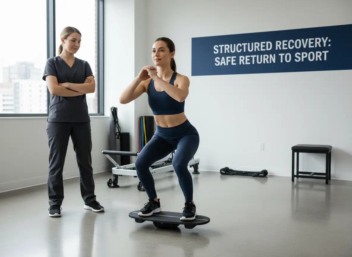
VRT is a customized exercise-based therapy that helps the brain adapt to vestibular dysfunction through habituation, adaptation, and substitution strategies. This therapy can significantly improve balance, reduce dizziness, and enhance daily function.
Anxiety and depression are common in patients with vestibular disorders, particularly those experiencing phobic postural vertigo or fear of falling. Studies show that anxious individuals are 4.65 times more likely to experience worsening vertigo, while those with depression have a 3.49 times greater risk. CBT helps manage psychological distress, improve gaze control, and reduce fear-related postural instability.
Medications can provide temporary relief from vestibular symptoms:
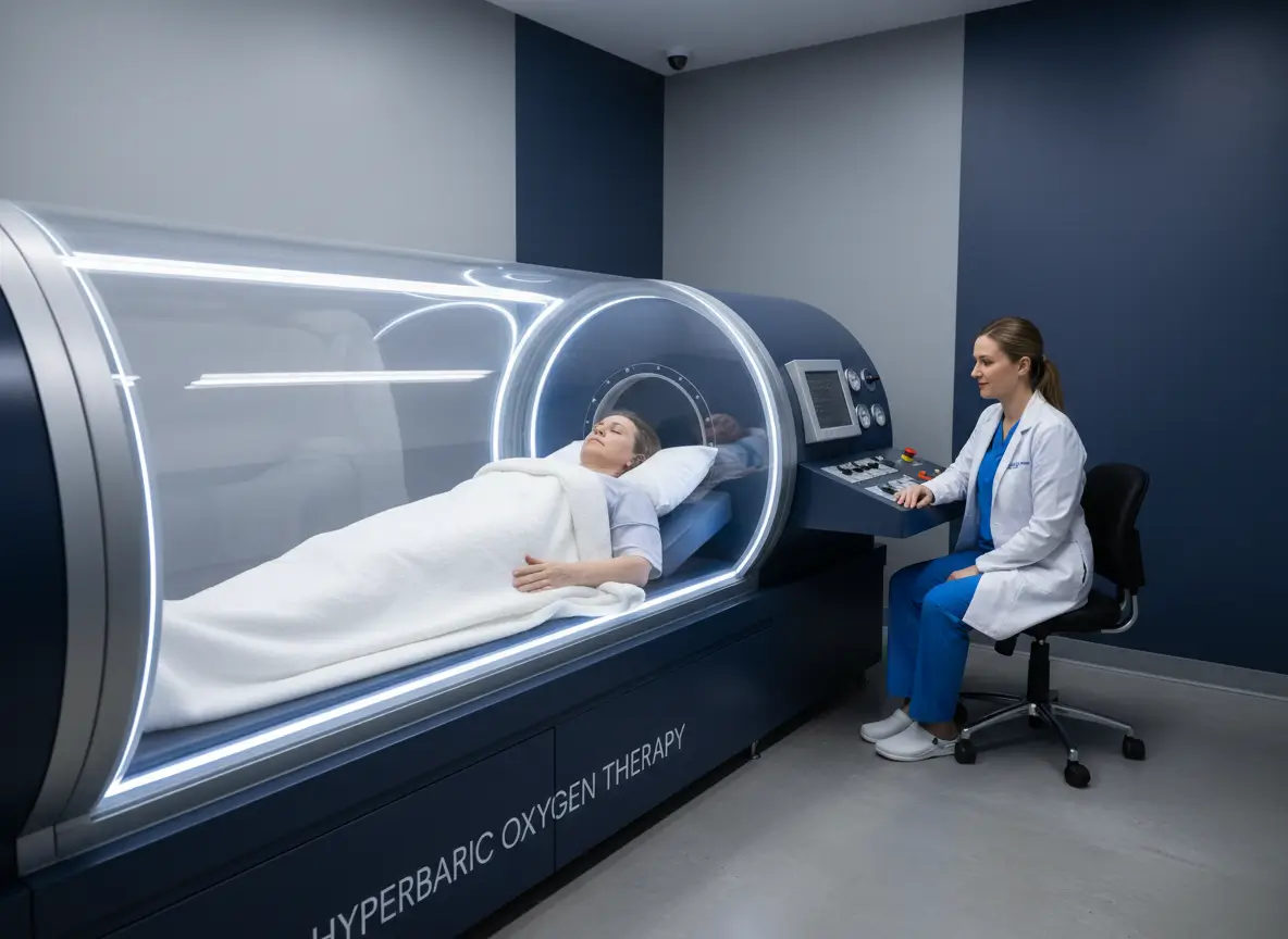
In severe cases, surgical procedures may be required:
Certain medications can damage the vestibular system, including aminoglycoside antibiotics (e.g., gentamicin), loop diuretics (e.g., furosemide), chemotherapy drugs (e.g., cisplatin), and high doses of aspirin (salicylates). Identifying and discontinuing these drugs, when possible, can prevent further vestibular damage.

Peripheral vestibular disorders encompass a broad spectrum of conditions, ranging from benign, self-limiting pathologies to progressive and debilitating disorders. Therefore, an accurate diagnosis and effective management require a multidisciplinary approach, integrating clinical evaluation, diagnostic testing, rehabilitation therapy, psychological support, and lifestyle modifications.
While some conditions can be permanently resolved, others necessitate long-term symptom management to enhance quality of life and functional independence.
At University Orthopedic Care, our multidisciplinary team consists of board-certified professionals in Neurosurgery, Orthopedic Surgery, Neuropsychology, and Physiatry (Physical Medicine and Rehabilitation).
We offer comprehensive vestibular evaluations and diagnostic tests, including:
For expert evaluation and personalized care, please do not hesitate to contact us at (866) 961-1744 or conveniently request an appointment online at University Orthopedic Care.